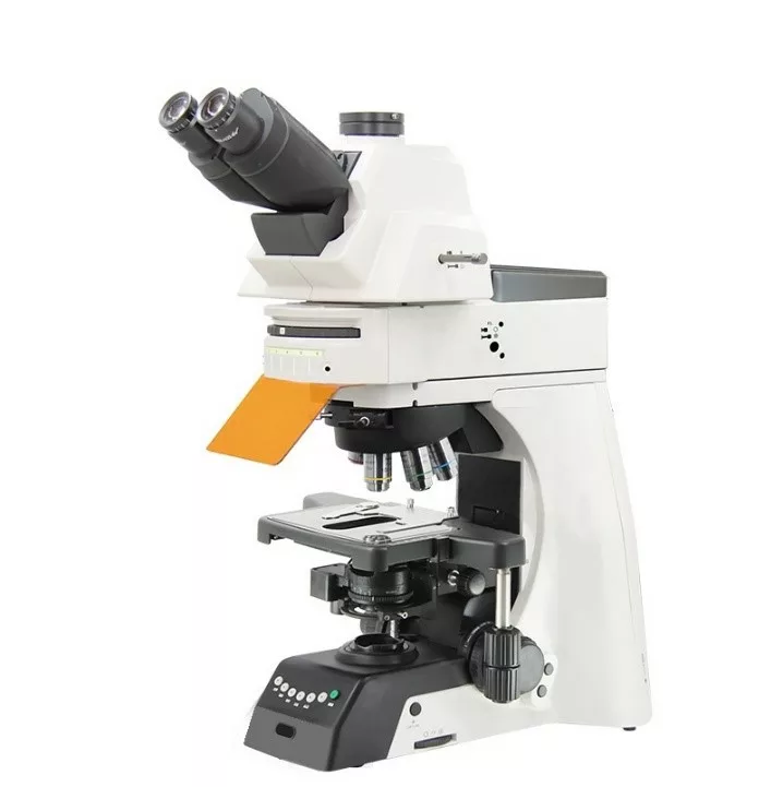
Fluorescent Microscope (Upright): Illuminating the World of Cellular and Molecular Biology
In the realms of cellular and molecular biology, the ability to visualize structures at the microscopic level is crucial. A Fluorescent Microscope KFM/U/LL stands out as an essential tool for researchers, allowing them to study biological samples with remarkable precision and clarity. By utilizing fluorescence, these microscopes enable scientists to observe specific cellular components, enhancing our understanding of complex biological processes and paving the way for groundbreaking discoveries.
What is a Fluorescent Microscope?
A Fluorescent Microscope is a specialized optical instrument designed to illuminate specimens with specific wavelengths of light, causing fluorescent dyes or proteins within the samples to emit light of a different wavelength. This unique property allows researchers to visualize specific cellular structures or molecules that would otherwise be difficult to detect with traditional brightfield microscopes. Fluorescent microscopy is widely used in various fields, including microbiology, immunology, and genetics.
How Does a Fluorescent Microscope Work?
Fluorescent microscopes utilize several key components to achieve high-resolution imaging:
- Light Source: High-intensity light sources, such as mercury or xenon lamps, emit short-wavelength light that excites fluorescent dyes within the specimen.
- Excitation Filter: This filter selects the specific wavelength of light needed to excite the fluorescent dye or protein in the sample.
- Objective Lenses: High-quality objective lenses focus the emitted light, magnifying the fluorescent signal while minimizing background noise.
- Emission Filter: This filter allows only the emitted fluorescence to pass through to the eyepiece or camera, blocking the excitation light and enhancing contrast.
- Detector: A camera or photomultiplier tube captures the emitted light, enabling researchers to visualize and analyze the fluorescent signal.
By combining these elements, fluorescent microscopes provide detailed images that reveal the intricate details of cellular structures.
Key Features of a High-Quality Fluorescent Microscope
When selecting a Fluorescent Microscope, consider the following essential features:
- Multicolor Imaging: Many advanced models allow for the simultaneous observation of multiple fluorescent markers, enabling complex experiments and analyses.
- High Resolution: Superior optical components and advanced imaging techniques ensure high-resolution images, revealing fine details in cellular structures.
- Ease of Use: User-friendly interfaces and controls make it accessible for researchers of all levels, facilitating smooth operation.
- Versatility: Adaptability for various applications, including live-cell imaging, fluorescence in situ hybridization (FISH), and immunofluorescence.
- Data Acquisition and Analysis: Integrated software for capturing, analyzing, and processing images enhances research efficiency and data management.
- Modularity: Many models offer modular designs that allow for easy upgrades and integration with additional imaging techniques, such as confocal microscopy.
Applications of Fluorescent Microscopes
Fluorescent microscopes are widely used in various scientific disciplines, including:
- Cell Biology: Essential for studying cellular processes, such as cell division, apoptosis, and signaling pathways.
- Molecular Biology: Used for techniques like FISH and immunofluorescence to visualize specific nucleic acids or proteins within cells.
- Microbiology: Allows researchers to observe the morphology and behavior of bacteria and other microorganisms through fluorescent staining.
- Neuroscience: Employed to visualize neurons and their connections, aiding in the understanding of brain function and pathology.
- Pathology: Vital for diagnosing diseases by detecting specific biomarkers in tissue samples, improving diagnostic accuracy.
Benefits of Using a Fluorescent Microscope
- Enhanced Visualization: Fluorescent microscopy provides high contrast and sensitivity, allowing for the observation of structures that are challenging to see with standard microscopy.
- Specificity: The ability to use different fluorescent markers enables targeted visualization of specific proteins or cellular components, facilitating detailed studies.
- Real-Time Imaging: Many fluorescent microscopes are capable of live-cell imaging, allowing researchers to observe dynamic biological processes as they unfold.
- Increased Productivity: Enhanced imaging capabilities lead to more efficient experiments and quicker results, accelerating the pace of research.
- Versatile Research Applications: Adaptable for various techniques, fluorescent microscopes cater to the diverse needs of researchers in multiple fields.
Choosing the Right Fluorescent Microscope for Your Needs
When selecting a Fluorescent Microscope, consider your specific research requirements, including the types of samples you will be studying and the necessary imaging capabilities. Evaluate factors such as optical quality, light source options, and the compatibility of fluorescent dyes or proteins you plan to use. Investing in the right microscope can significantly enhance your research outcomes.
Conclusion
A Fluorescent Microscope is an indispensable tool for researchers in cellular and molecular biology, enabling the visualization of intricate cellular structures and processes with exceptional clarity. By harnessing the power of fluorescence, these microscopes facilitate groundbreaking research, advancing our understanding of biology and contributing to significant scientific discoveries. Equip your laboratory with a high-quality fluorescent microscope to unlock the full potential of your research.


Reviews
There are no reviews yet.